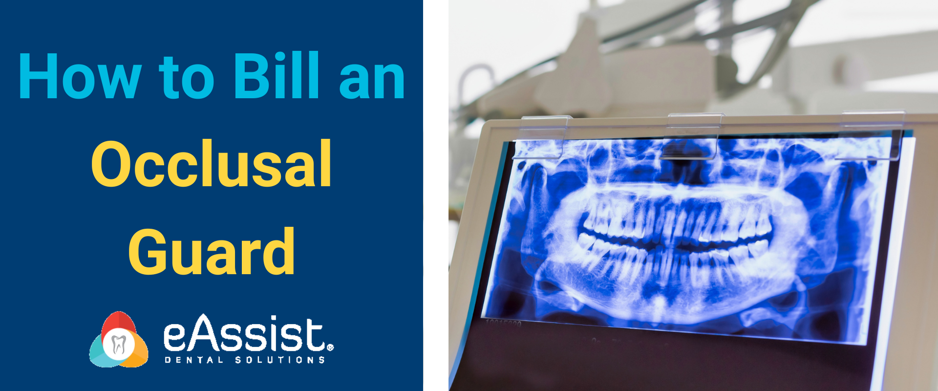
Quality radiographs are essential for complete diagnosis. While visual detection is important many conditions can only be seen by radiograph such as lesions on the apex of the tooth or changes in pulpal health and evidence of loss of supportive bone structure.
When filing claims for the dental practice, it has been a challenge many a time to find a good radiograph that documents the condition of the tooth related to the procedure performed.
If the root tip is cut off or there is overlapping on the interproximal contacts it is impossible to prove that there is infection at the root tip or caries between the teeth.
One of the top film exposure errors is cutting off the root apices or tips on films. The cause is improper positions of placement often because it is uncomfortable for the patient and there is movement at exposure. The solution would be to reposition the film, receptor or PSP closer to the midline where the palate and floor of the mouth are deeper. Increase the vertical angulation (not more than 15 degrees from the perpendicular) will help prevent cutting off the root tips. It is helpful to use a Rinn XCP or BAI which helps eliminate the guesswork.
The solution to overlapping a bitewing exposure is to recheck the angle of the beam that it is not too mesial or distal to the interproximal. Adjust the horizontal angulation by moving the tube head side to side. You then reposition the film using a XCP, Snap-a-ray or Bitewing tab so that it is parallel to the facial surfaces of the teeth being exposed. Adjust the horizontal angulation so that the central ray is aimed at the open contacts of the teeth being x-rayed. You may have to adjust the vertical angle by 5 to 10 degrees up or down of the tube head to get the beam at the interproximal.
If you are using regular film be careful not to bend it to get it into the patient’s mouth as this will cause a distortion. Cone cutting is another common exposure error caused by lack of x-ray exposure; the beam is not centered in the target area.
There are many more problems associated with creating great films and practices should have quality control in place to assure that all operators are trained to take diagnostic films with safety for the patient at the core. Even with the use of digital x-rays, patients do not want to be over exposed because the operator is not trained in technique.
Many an insurance claim has been denied due to poor diagnostic x-rays needed to validate the payment of the claim. The clinical team must be trained to ensure that excellent diagnostic and relevant radiographs are provided with every restorative, periodontal, endodontic and oral surgery procedure.
It is the dentist’s responsibility to order the proper radiographs and to interpret them for quality while the patient is in the treatment room and prior to services rendered.
For compliance, for procedure verification, for claims management and for patient/customer service, diagnostic radiographs are necessary for practice success.


0 Comments